Scoliosis in Infants

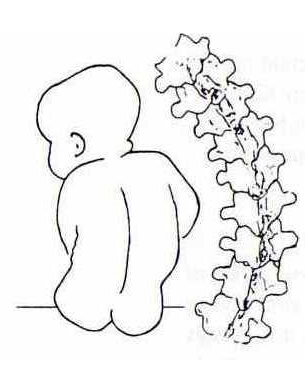
Early Onset scoliosis or Infantile Idiopathic scoliosis is defined as scoliosis that is first diagnosed in a child between birth and 5 years of age. Parents usually first notice that their child persistently lies in a “banana shape” or sits leaning to one side. Also, a prominence of the ribs on one side of the back or chest may be seen or felt. Scoliosis in infants usually resolve without treatment but the ones that do not resolve (approx. 10%) can be difficult to manage. A magnetic resonance imaging (MRI) is often used to determine if there are any abnormalities of the spinal cord or spinal column. Sedation or even general anesthesia may be used to relax the child enough to obtain quality images. Many infantile curves are left-sided curves in the thoracic (middle) spine, where as they occur more  commonly on the right side in adolescent idiopathic scoliosis. Many infants are otherwise healthy and just have a small curvature of the spine. In some infants there is an increased association with hip dysplasia, mental retardation, and congenital heart disease.
Congenital scoliosis (caused by malformed and/or connected vertebrae) is also diagnosed during this period. However, these curves are not included in the infantile idiopathic scoliosis category. Congenital scoliosis forms during the first six weeks of embryonic formation.
Scoliosis fusion surgery is not used in scoliosis in infants because the spine and lungs of an infant are not mature and they need to fully develop in size. A fusion would prevent further growth of the instrumented segment of the spine. Also, depending on the type of instrumentation used, the anterior spine may continue to grow leading to “crankshaft phenomenon”. This occurs when the front part (anterior) of the spine is fused from the back (posterior) and the spine twists as it grows. In this situation, the infantile scoliosis continues to progress despite the posterior fusion. For this reason, other treatments have been developed for the management of early onset scoliosis. These techniques take into account the growth of the spine as well as the growth of the rib cage and lungs. If implants are prescribed, multiple expansions or lengthenings may be necessary (usually twice per year) to keep up with growth in the young spine.
It is typically advised that parents wait 3-6 months to determine whether or not the scoliosis will progress. However, there is a measuring technique used with scoliosis in infants to help determine if the curve is progressive in nature or self resolving. The Rib Vertebral Angle Degree (RVAD) is measured and analyzed to give an indication of what type of curve is present. Because maximum correction and curve resolution depend on the time in which treatment is administered, the sooner treatment is started the better and the greater the chance of success.
Early treatment with a serial corrective plaster Elongation Derotation Flexion (EDF) scoliosis cast is conducted as soon as the infantile scoliosis is considered progressive. The infant’s curve will keep pace at the rate in which the child is growing, which translates to 24 cm.s in the first two years. Plaster of Paris is typically used in the application of the scoliosis cast. This is because plaster doesn’t dry as fast as fiberglass and therefore allows the time necessary to address rotation and apply the most effective scoliosis cast. A layer of fiberglass is applied on the outside of the scoliosis cast to protect the plaster.
A large cutout in the front of the scoliosis cast allows the child breathing room and provides rib flaps to support the rib cage and prevent permanent rib flaring. There is another cutout in the back that is trimmed on the concavity side of the scoliosis curve. This cutout allows the flattened ribs on the concave side of the curve to grow out and the prominent ribs on the convex side to grow flat.
For more information please visit:
