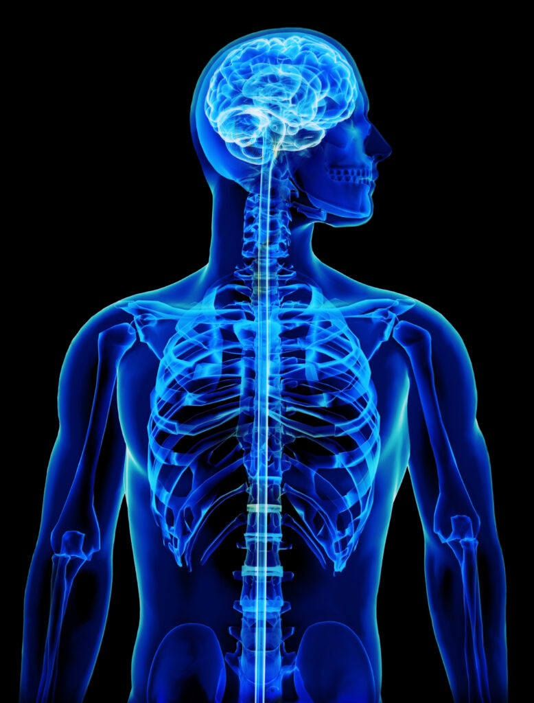
*RESEARCH VIDEO (Patients do not receive DMX as standard treatment protocol) DMX or “Digital Motion X-ray” is a type of fleuro-based X-ray coupled with digital and optic technology which allows clinicians to view the spine in real time motion.
By subjecting all the therapies, exercises, and spinal manipulations to scrutiny under digital motion X-ray, Dr. Strauss is able to clarify effects on the scoliosis. In this video you are seeing Dr. Strauss getting his cervical spine manipulated with the Arthrostim instrument by a fellow doctor. This was part of a research and training project. His mouth is opened to allow better viewing of the upper cervical spine.
How can Digital Motion X-ray Provide Efficacy of Care Program?
Digital Motion X-ray (DMX) is a diagnostic imaging technique that combines real-time X-ray imaging with motion capture technology. It allows healthcare professionals to assess the movement and function of joints and other anatomical structures during motion. While DMX itself is a diagnostic tool rather than a program, it can be used within an Efficacy of Care program to enhance patient care and treatment outcomes. Here’s how DMX can contribute to such a program:
- Comprehensive evaluation: DMX provides a dynamic view of the body’s structures, enabling a more thorough evaluation of musculoskeletal conditions and injuries. By capturing X-ray images in real-time during specific movements or activities, it allows healthcare providers to identify issues that may not be visible in static images, such as conventional X-rays or MRI scans. This comprehensive evaluation can lead to more accurate diagnoses and treatment plans.
- Treatment planning: By visualizing joint motion and anatomical abnormalities in real-time, DMX helps healthcare providers better understand the underlying causes of pain, dysfunction, or limitations in movement. This information is valuable in developing personalized treatment plans that address the specific needs of each patient. The dynamic nature of DMX allows for precise targeting of treatment interventions and may help optimize outcomes.
- Monitoring progress: With Digital Motion X-ray, healthcare professionals can monitor patients’ progress during treatment and rehabilitation. By comparing sequential DMX scans, they can assess changes in joint function, alignment, or range of motion over time. This objective data provides valuable insights into the effectiveness of the chosen treatment strategies and helps healthcare providers make informed decisions regarding ongoing care and adjustments to treatment plans.
- Patient education and engagement: Digital Motion X-ray provides visual evidence of the patient’s condition, making it easier for healthcare providers to educate patients about their diagnosis, treatment options, and the importance of adhering to prescribed interventions. The real-time imaging during motion can help patients better understand their limitations and how their movements impact their condition. This increased understanding can lead to improved patient compliance and engagement in their own care, potentially enhancing treatment outcomes.
- Research and data analysis: Digital Motion X-ray can be a valuable tool for research purposes within an Efficacy of Care program. The dynamic imaging capabilities of DMX allow for the collection of quantitative data on joint movements, kinematics, and pathologies. This data can be analyzed to gain insights into treatment outcomes, efficacy of different interventions, and the progression of specific conditions. Such research findings can contribute to evidence-based practices and further refine treatment protocols.
In summary, Digital Motion X-ray can provide a range of benefits within an Efficacy of Care program by enabling comprehensive evaluations, aiding treatment planning, monitoring progress, enhancing patient education and engagement, and contributing to research and data analysis. By utilizing DMX alongside other diagnostic and therapeutic modalities, healthcare providers can optimize patient care and improve treatment efficacy.
Also read: Isometric & Stretching Exercises For Scoliosis
About:
Dr. Strauss is the director of the Hudson Valley Scoliosis Correction Center in New York. He has been actively engaged in scoliosis treatment for the past 30 years and has authored two books on the subject, Your Child Has Scoliosis and The Truth About Adult Scoliosis.
He is Vice President of the CLEAR Scoliosis Institute and a lecturer for their introductory and advanced workshops. He is certified in scoliosis bracing and in the use of scoliosis specific exercises. Dr. Strauss is a graduate of the ISICO World Masters of Scoliosis.His postgraduate studies also include a Masters Degree in Acupuncture as well as training in Grostic, Pettibon, CBP, Clinical Nutrition, Chinese Herbal Medicine, Manipulation under Anesthesia, and Electrodiagnosis.
His scoliosis practice has treated patients from 25 states and 32 other foreign countries.
If you have questions about childhood and adult scoliosis and how it can be successfully treated without surgery subscribe to our channel!
